Quantitative bone marrow MRI
Research summary
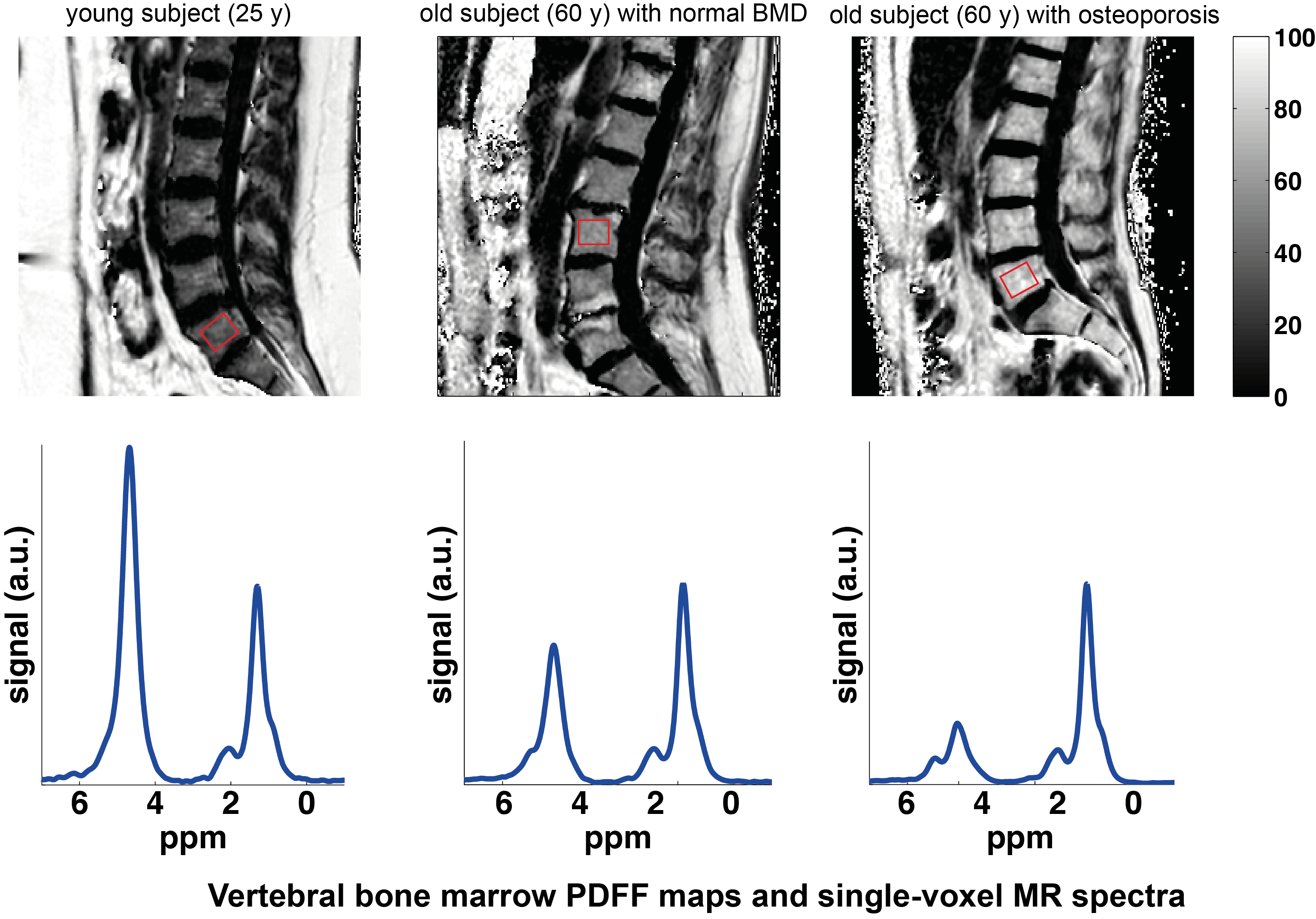
Osteoporosis has an estimated prevalence of up to 30% among post-menopausal women in Europe. Osteoporotic spine and hip fractures considerably reduce quality of life and are associated with an increased mortality. Osteoporosis can be treated using known medications, but its current early diagnosis based on bone mineral density (BMD) measurements remains insufficient and poorly correlated with fracture risk. In addition to the role of the bone mineral matrix, there is recent preliminary evidence that bone marrow fat plays a role in the pathophysiology of bone disease. MRI is the ideal tool to quantitatively measure properties of bone marrow and can be also used to simultaneously indirectly assess trabecular bone density.
Our bone and bone marrow MRI research focuses on the following directions:
- Addressing confounding effects in the measurement of the bone marrow proton density fat fraction (PDFF) using both single-voxel MRS and chemical shift encoding-based water–fat separation.
- Development of single-voxel MRS techniques to measure fat unsaturation in red bone marrow regions (e.g. spine).
- Development of quantitative susceptibility mapping (QSM) techniques to measure bone marrow magnetic susceptibility and investigate the relationship between bone marrow susceptibility, T2*, and bone mineral density.
- Application of the developed quantitative MRI and MRS techniques in clinical studies of patients with primary osteoposoris, secondary osteoporosis and diabetes, in order to improve bone loss diagnostics and pathophysiology understanding.
Collaborators
PD Dr. T. Baum, Dr. A. Gersing, PD Dr. J. Kirschke, Dr. B. Schwaiger, Prof. Rychlik (Food Chemistry)
Key publications
-
Dieckmeyer, M., Ruschke, S., Eggers, H., Kooijman, H., Rummeny, E.J., Kirschke, J.S., Baum, T., Karampinos, D.C., ADC Quantification of the Vertebral Bone Marrow Water Component: Removing the Confounding Effect of Residual Fat. Magnetic Resonance in Medicine 78, 1432–1441.
-
Cordes, C., Baum, T., Dieckmeyer, M., Ruschke, S., Diefenbach, M.N., Hauner, H., Kirschke, J.S., Karampinos, D.C., MR-Based Assessment of Bone Marrow Fat in Osteoporosis, Diabetes, and Obesity. Frontiers in Endocrinology 7, 74.
-
Karampinos, D.C., Ruschke, S., Dieckmeyer, M., Eggers, H., Kooijman, H., Rummeny, E.J., Bauer, J.S., Baum, T., Modeling of T 2 * Decay in Vertebral Bone Marrow Fat Quantification. NMR in Biomedicine 28, 1535–1542.
-
Baum, T., Yap, S.P., Dieckmeyer, M., Ruschke, S., Eggers, H., Kooijman, H., Rummeny, E.J., Bauer, J.S., Karampinos, D.C., Assessment of Whole Spine Vertebral Bone Marrow Fat Using Chemical Shift-Encoding Based Water-Fat MRI. Journal of Magnetic Resonance Imaging 42, 1018–1023.
-
Baum, T., Inhuber, S., Dieckmeyer, M., Cordes, C., Ruschke, S., Klupp, E., Jungmann, P.M., Farlock, R., Eggers, H., Kooijman, H., Rummeny, E.J., Schwirtz, A., Kirschke, J.S., Karampinos, D.C., Association of Quadriceps Muscle Fat With Isometric Strength Measurements in Healthy Males Using Chemical Shift Encoding-Based Water-Fat Magnetic Resonance Imaging. Journal of Computer Assisted Tomography 40, 447–451.
-
Dieckmeyer, M., Ruschke, S., Cordes, C., Yap, S.P., Kooijman, H., Hauner, H., Rummeny, E.J., Bauer, J.S., Baum, T., Karampinos, D.C., The Need for T2 Correction on MRS-Based Vertebral Bone Marrow Fat Quantification: Implications for Bone Marrow Fat Fraction Age Dependence. NMR in Biomedicine 28, 432–439.
-
Karampinos, D.C., Melkus, G., Baum, T., Bauer, J.S., Rummeny, E.J., Krug, R., Bone Marrow Fat Quantification in the Presence of Trabecular Bone: Initial Comparison Between Water-Fat Imaging and Single-Voxel MRS. Magnetic Resonance in Medicine 71, 1158–1165.
-
Baum, T., Yap, S.P., Karampinos, D.C., Nardo, L., Kuo, D., Burghardt, A.J., Masharani, U.B., Schwartz, A.V., Li, X., Link, T.M., Does Vertebral Bone Marrow Fat Content Correlate With Abdominal Adipose Tissue, Lumbar Spine Bone Mineral Density, and Blood Biomarkers in Women With Type 2 Diabetes Mellitus? Journal of Magnetic Resonance Imaging 35, 117–124.