Quantitative fat MRI in metabolic syndrome
Research summary
The metabolic syndrome is a cluster of conditions, including obesity and insulin resistance, that increases by two-fold the risk for developing cardiovascular disease (CVD) and by five-fold the risk for developing type 2 diabetes. The prevalence of the metabolic syndrome is estimated between 20% and 30% in the adult population of most countries. The selection of optimal strategies for preventing diabetes and CVD for each individual patient is problematic using currently available biomarkers. Quantitative MRI of different fat depots can assist in improving the metabolic phenotyping of patients with metabolic syndrome with the ultimate goal to improve the selection of optimal prevention strategies for each individual patient.
Our fat MRI research focuses on the following directions:
- Development of quantitative MRI biomarkers to quantify fat content and fatty acid composition in different organs (liver, skeletal muscle, adipose tissue).
- Development of diffusion MRS and MRI techniques to study diffusion properties of adipose tissue.
- Development of non-invasive techniques to differentiate between brown and white adipose tissue.
- Development of efficient algorithms for the segmentation of the different abdominal fat depots.
- Application of the developed techniques in longitudinal lifestyle intervention studies involving diet and physical exercise.
Collaborators
Prof. H. Hauner (Nutrition), Prof. B. Menze (Computer Science), Prof. V. Ntziachristos (Optical Imaging)
Key publications
-
Shen, J., Baum, T., Cordes, C., Ott, B., Skurk, T., Kooijman, H., Rummeny, E.J., Hauner, H., Menze, B.H., Karampinos, D.C., Automatic Segmentation of Abdominal Organs and Adipose Tissue Compartments in Water-Fat MRI: Application To Weight-Loss in Obesity. European Journal of Radiology 85, 1613–1621.
-
Baum, T., Cordes, C., Dieckmeyer, M., Ruschke, S., Franz, D., Hauner, H., Kirschke, J.S., Karampinos, D.C., MR-Based Assessment of Body Fat Distribution and Characteristics. European Journal of Radiology 85, 1512–1518.
-
Franz, D., Karampinos, D.C., Rummeny, E.J., Souvatzoglou, M., Beer, A.J., Nekolla, S.G., Schwaiger, M., Eiber, M., Discrimination Between Brown and White Adipose Tissue Using a two-point Dixon Water-Fat Separation Method in Simultaneous PET/MRI. Journal of Nuclear Medicine 56, 1742–1747.
-
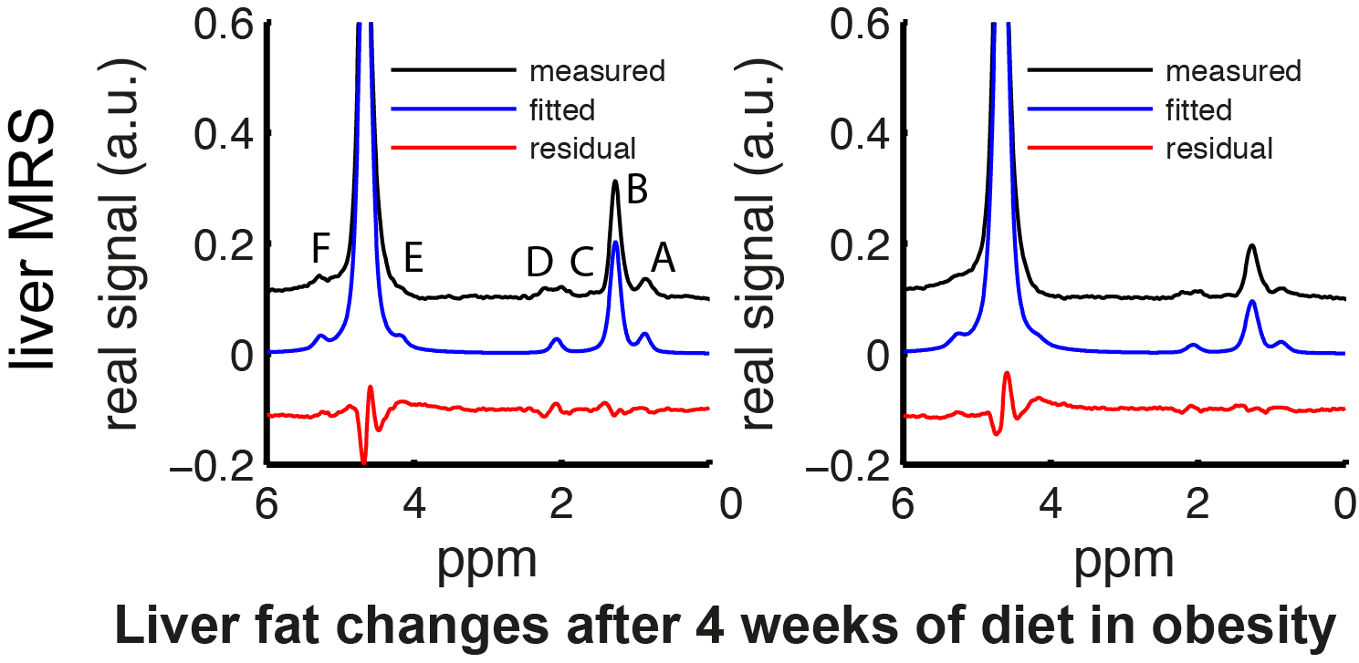
Cordes, C., Dieckmeyer, M., Ott, B., Shen, J., Ruschke, S., Settles, M., Eichhorn, C., Bauer, J.S., Kooijman, H., Rummeny, E.J., Skurk, T., Baum, T., Hauner, H., Karampinos, D.C., MR-Detected Changes in Liver Fat, Abdominal Fat, and Vertebral Bone Marrow Fat After a Four-Week Calorie Restriction in Obese Women. Journal of Magnetic Resonance Imaging 42, 1272–1280.
-
Karampinos, D.C., Baum, T., Nardo, L., Alizai, H., Yu, H., Carballido-Gamio, J., Yap, S.P., Shimakawa, A., Link, T.M., Majumdar, S., Characterization of the Regional Distribution of Skeletal Muscle Adipose Tissue in Type 2 Diabetes Using Chemical Shift-Based Water–fat Separation. Journal of Magnetic Resonance Imaging 35, 899–907.