MR Neurography
Research summary
Peripheral neuropathy describes damage to the nerves of the peripheral nervous system with primary causes being trauma, tumors or the side effects of systemic disease (e.g. diabetes). It is estimated that about 5% of population has some form of neuropathy and there is up to 5% incidence of peripheral nerve injuries in admissions to a level I trauma center. Furthermore, according to recent studies, approximately half of all patients with type II diabetes mellitus develop diabetic neuropathy. Clinical evaluation of peripheral neuropathies has traditionally relied on clinical examination and electrodiagnostic testing. However, these methods can frequently provide incomplete information about the anatomy and degree of peripheral neuropathy. MR neurographic techniques enable the non-invasive morphological and functional imaging of the peripheral nervous system.
Our peripheral nerves MRI research focuses on the following directions:
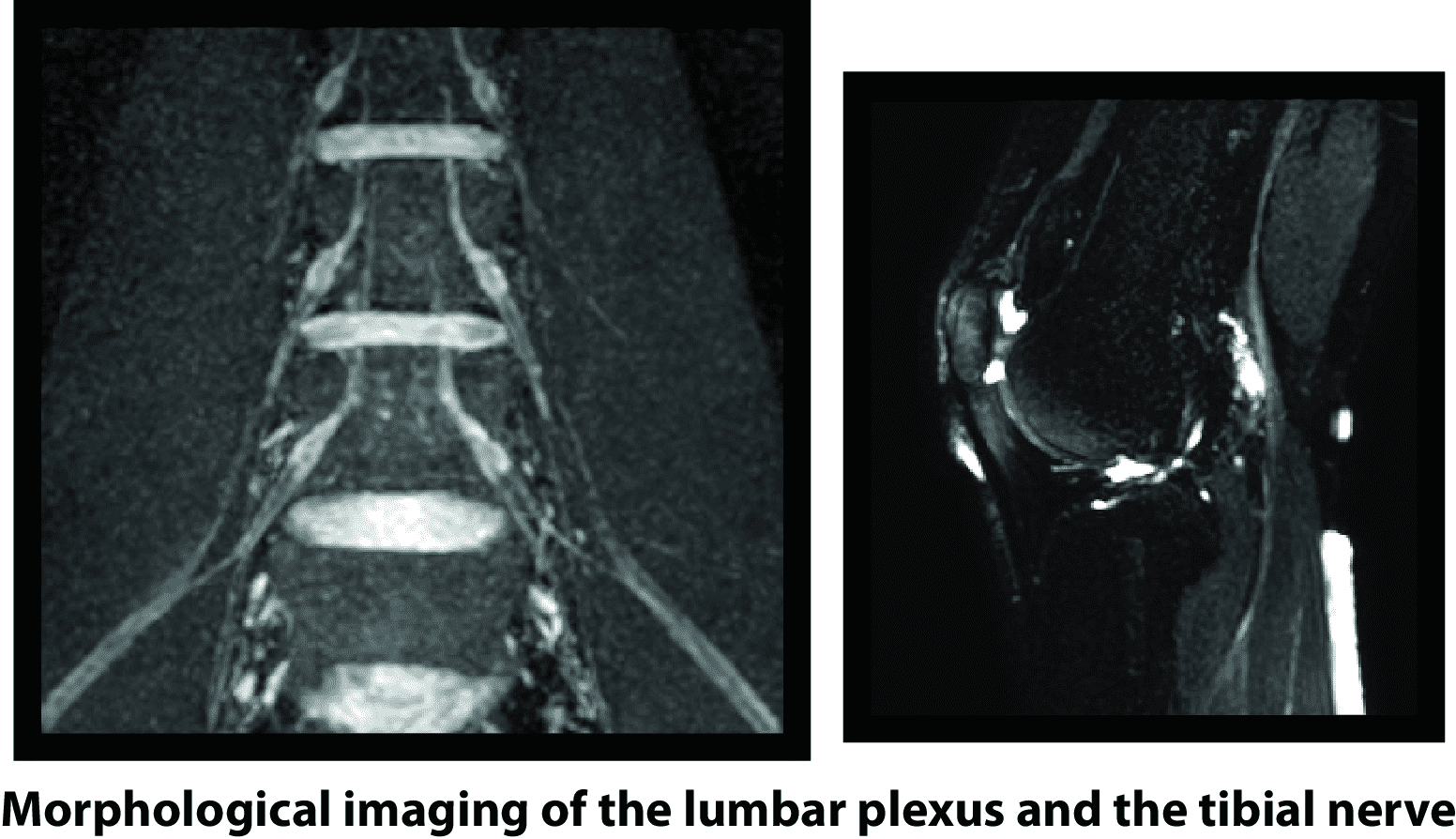
- Development of high-resolution three-dimensional neurographic imaging methods aiming to provide good nerves delineation in the lumbar plexus, the brachial plexus and the extremities, suppressing the contaminating effects of surrounding signals from vessels, fat and CSF.
- Development of high-isotopic resolution T2 mapping and DTI methods for imaging the long axis of peripheral nerves in the extremities and the lumbar plexus.
- Application of the developed techniques in human volunteer studies to establish normative peripheral nerve T2 and DTI values and in clincal studies of patients with nerve entrapements and neurogenic tumors in order to characterize axonal degeneration/regeneration and to visualize nerve tracts.
Affiliated members
Nico Sollmann, Elisabeth Klupp
Collaborators
PD Dr. J. Kirschke, Prof. K. Woertler, Dr. A. Gersing
Key publications
-
Cervantes, B., Kirschke, J.S., Klupp, E., Kooijman, H., Börnert, P., Haase, A., Rummeny, E.J., Karampinos, D.C., Orthogonally combined motion- and diffusion-sensitized driven equilibrium (OC-MDSDE) preparation for vessel signal suppression in 3D turbo spin echo imaging of peripheral nerves in the extremities. Magnetic Resonance in Medicine 79, 407–415.
-
Cervantes, B., Bauer, J.S., Zibold, F., Kooijman, H., Settles, M., Haase, A., Rummeny, E.J., Wörtler, K., Karampinos, D.C., Imaging of the Lumbar Plexus: Optimized Refocusing Flip Angle Train Design for 3d TSE. Journal of Magnetic Resonance Imaging 43, 789–799.
-
Karampinos, D.C., Melkus, G., Shepherd, T.M., Banerjee, S., Saritas, E.U., Shankaranarayanan, A., Hess, C.P., Link, T.M., Dillon, W.P., Majumdar, S., Diffusion Tensor Imaging and T 2 Relaxometry of Bilateral Lumbar Nerve Roots: Feasibility of In-Plane Imaging. NMR in Biomedicine 26, 630–637.